Our Office
1941 Coney Island Avenue
Brooklyn, NY 11223
Existing Patients: (718) 400-2396
New Patients: (718) 412-9457
Fax: (718) 400-8932
Visit Us Online

Digital radiography replaces traditional film with sensitive electronic sensors and computer software to capture dental images. Instead of waiting for film to develop, the image appears on a monitor almost instantly, allowing your child’s dental team to review what they see right away. This shift from chemical processing to digital capture has reshaped how pediatric dental care is delivered—making visits more efficient and information more accessible.
One of the biggest practical differences is how images are recorded and handled. Digital sensors come in a few sizes and forms tailored for pediatric use, and the picture files are stored immediately in the patient’s electronic chart. That means fewer delays between taking an image and planning treatment, as well as the ability to compare current images with previous ones over time without handling fragile film sheets.
For parents, the change is reassuring: digital systems typically expose children to lower doses of radiation than conventional film x-rays while producing clearer images for diagnosis. The combination of reduced exposure and faster results makes digital radiography particularly well suited to the needs and sensitivities of young patients.
Digital images can be enhanced with software tools that adjust contrast, brightness, and zoom so the dentist can see subtle details that might be missed on film. These enhancements do not alter the underlying information; they simply make it easier for clinicians to detect early signs of decay, monitor root development, or evaluate trauma. Clearer visualization supports more confident clinical decisions and targeted treatment plans.
Because images are available instantly, the dental team can discuss findings with parents during the same visit. This real-time review encourages collaborative decision-making: clinicians can point out areas of concern on the screen, explain why a particular image was taken, and outline next steps in plain language. Immediate access also reduces the need for return appointments solely for diagnostic follow-up.
Digital files are easy to share securely with other healthcare providers when a referral or collaborative opinion is needed. Multiple practitioners can view the same high-resolution image without the physical transfer of film, which streamlines consultations and helps ensure continuity of care across specialties when complex issues arise.
Modern digital sensors are designed with pediatric comfort in mind. Smaller sensor sizes and flexible positioning options help minimize discomfort during image capture, an important consideration when working with infants, toddlers, and anxious children. Our staff are trained to use gentle techniques that reduce movement and shorten the time a sensor needs to remain in the child’s mouth.
Digital radiography also contributes to safety in two meaningful ways. First, the exposure levels used are typically lower than with conventional film, which reduces cumulative radiation over a child’s lifetime. Second, the speed and accuracy of digital imaging reduce the likelihood of repeat exposures caused by improper technique or indistinct images, further limiting unnecessary radiation.
Infection control practices remain a top priority when using any intraoral device. Sensors are protected with barriers and cleaned according to strict protocols between patients. Combining careful handling, protective coverings, and digital technology helps create an environment that is both safe and reassuring for families.
Digital radiography plays a central role across many aspects of pediatric dentistry, from routine checkups to treatment planning. During preventive visits, bitewing or periapical images can reveal early cavities between teeth or under existing restorations long before they become visible on a clinical exam. Early detection allows for less invasive, more conservative care focused on preserving healthy tooth structure.
For developmental monitoring, digital images provide a reliable way to track how permanent teeth are erupting and how roots are forming. This information is important for timing interventions such as space maintainers or guidance for incoming teeth. In cases of injury or dental trauma, rapid digital imaging helps the team assess the extent of damage and determine whether urgent treatment is needed.
When restorative procedures are indicated, digital radiographs assist in precise treatment planning—helping to size crowns, evaluate the fit of restorations, and confirm completion of certain procedures. The ability to instantly review the outcome of a treatment with parents present builds understanding and trust without relying on delayed film development or follow-up calls.
Because digital images can be archived and compared over time, they are also a valuable educational tool. Parents can visually track progress, understand the reasons behind recommended preventive measures, and see improvements following treatment—information that supports long-term oral health habits for children.
Digital radiography integrates directly with electronic health records, ensuring that every image becomes part of the child’s secure dental chart. Images are stored with appropriate access controls and backups, which helps protect patient privacy while maintaining reliable records for future reference. This streamlined record keeping reduces paperwork and makes it easier to retrieve images when coordinating care with schools, specialists, or other providers when appropriate.
From an environmental perspective, removing film development from the process eliminates the need for chemical developers and fixers, along with the associated disposal issues. That makes digital imaging a cleaner option for practices aiming to reduce their environmental footprint while maintaining high standards of care.
Ongoing software updates and staff training ensure that the technology continues to perform optimally. Quality assurance measures—such as routine equipment calibration and image audits—help maintain consistent image clarity and diagnostic reliability. Together, these practices create a dependable digital imaging workflow that supports excellent pediatric dental care.
Digital radiography is a child-centered approach to dental imaging that combines lower radiation exposure, faster results, and clearer diagnostic images with improved comfort and safer infection control. In the hands of a trained pediatric dental team, these advantages translate into earlier detection, more precise treatment planning, and a more positive experience for families.
If you’d like to learn more about how we use digital x-rays for children at Beyond Smiles Pediatric Dentistry or have questions about safety and imaging protocols for your child, please contact us for more information.
Digital radiography uses electronic sensors and computer software to capture dental images instead of film and chemical processing. Images appear on a monitor almost instantly, eliminating the wait for film development and allowing clinicians to review results during the same visit. Because files are stored digitally, clinicians can compare current images with prior studies without handling fragile film sheets.
Unlike film, digital systems allow for on-screen enhancements such as adjusted contrast, brightness, and zoom to help identify subtle problems. These tools do not change the diagnostic information; they make important details easier to see. The digital workflow also streamlines archiving and retrieval, which improves record keeping and care coordination.
Digital radiography is well suited to pediatric dentistry because sensors come in smaller sizes and flexible designs that improve fit and comfort for infants, toddlers, and school-age children. The typically lower radiation doses used with digital detectors reduce cumulative exposure over time while still producing high-resolution images for accurate diagnosis. Faster image capture and immediate review shorten appointments and reduce the need for repeat exposures caused by indistinct film.
For families, digital images also support clearer communication: clinicians can review images with parents during the appointment and explain concerns using the image on-screen. This real-time approach helps parents understand findings and the reasoning behind recommended preventive or restorative steps. It also reduces anxiety when clinicians can show rather than only describe what they are seeing.
Digital sensors produce high-resolution images that can be enhanced with software to reveal fine details such as early decay, root development, or trauma-related changes. Tools that adjust contrast, brightness, and magnification help clinicians detect issues that might be harder to see on conventional film. These enhancements support more confident treatment decisions without altering the underlying image data.
Because images are instantly available, clinicians can evaluate results immediately and, if necessary, take additional views while the child is still in the chair. Routine quality checks and periodic equipment calibration further ensure consistency and reliability of the images over time. Together, these capabilities reduce diagnostic uncertainty and support precise treatment planning.
When used appropriately, digital x-rays are considered safe for pediatric patients because they typically require lower radiation doses than conventional film radiography. Protective measures such as lead aprons, thyroid collars when indicated, and adherence to pediatric imaging protocols limit exposure and follow the principle of keeping doses as low as reasonably achievable. Staff training and careful technique also reduce the chance of repeat images, which helps minimize cumulative exposure.
Decisions about whether and how often to take radiographs are individualized based on a child’s age, dental development, risk of disease, and clinical findings. Clinicians follow established guidelines to balance the diagnostic benefits of imaging against the goal of minimizing radiation. Parents are encouraged to ask about safety measures and the specific reasons an image is recommended for their child.
Modern digital sensors are designed with pediatric comfort in mind, with smaller sizes and flexible positioning that reduce the amount of time a device needs to stay in the mouth. Staff use gentle positioning techniques, clear, age-appropriate instructions, and distraction strategies to help children remain calm and still during image capture. Shorter exposure times and instant image review also decrease repeat attempts, which reduces both discomfort and anxiety.
For very young or anxious children, the team may use additional behavior guidance methods and supportive communication to create a positive experience. Preparing the child with a simple explanation and allowing a parent to be nearby when appropriate helps many children feel more secure. These approaches support cooperation while maintaining safe and effective imaging practice.
The frequency of dental x-rays is based on each child’s individual needs, including age, oral health status, risk of decay, and the stage of dental development. Routine bitewing or periapical images may be taken at periodic intervals to detect early cavities between teeth or under restorations that are not visible during a clinical exam. Developmental images are scheduled as needed to monitor eruption patterns and root formation, which can guide interventions such as space maintainers.
Emergency or trauma situations may require immediate imaging to assess damage and plan urgent care. Your child’s pediatric dental team will explain the clinical reasons for any recommended images and tailor the schedule to minimize exposure while ensuring timely, accurate diagnosis. Parents should feel free to discuss imaging plans and any concerns with the care team.
Digital radiographs are integrated directly into the electronic dental record, where they are stored with access controls, backups, and audit trails to protect patient privacy. Secure storage makes it easier to retrieve images for future reference, compare studies over time, and include them in comprehensive treatment records. This centralization also reduces paperwork and the risk of losing or misplacing film.
Access to images is limited to authorized members of the dental team and, when appropriate, other health professionals involved in the child’s care. Practices follow data security protocols and routine system maintenance to safeguard electronic records. Parents can ask about record retention policies and the measures taken to protect their child’s health information.
Yes, digital images are easily and securely shared with other dental specialists, pediatricians, or healthcare providers when a referral or collaborative opinion is needed. High-resolution files can be transmitted electronically without the physical transfer of film, which streamlines consultations and helps ensure continuity of care across providers. Secure sharing protocols protect patient privacy during the transfer process.
Sharing images facilitates coordinated treatment planning for complex cases, including orthodontic evaluation, surgical referrals, or multidisciplinary management after trauma. When a referral is made, the dental team typically includes the relevant images and clinical notes to help the receiving provider understand the clinical context. Parents are informed about what will be shared and why.
Digital radiographs allow early detection of decay between teeth and under restorations, making it possible to treat problems conservatively before they progress. For preventive care, images help clinicians identify high-risk areas and monitor the effect of preventive measures such as fluoride or sealants. When restorative treatment is necessary, digital images assist in precise planning, sizing restorations, and checking fit and completion of procedures.
Because images can be archived and compared over time, they serve as an educational tool for families to visualize progress and the impact of recommended habits. Immediate review of post-treatment images also helps parents understand the outcome and supports informed follow-up care. This visual feedback encourages adherence to preventive strategies that protect long-term oral health.
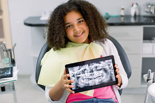
During a visit, the team will explain why a radiograph is recommended and describe the steps they will take to keep your child comfortable and safe. A small digital sensor will be positioned briefly in the child’s mouth and the image will be captured in seconds, then reviewed on-screen with you and your child. Protective measures such as a lead apron and careful technique are used when indicated to reduce exposure.
After imaging, clinicians will discuss any findings and recommended next steps in plain language, showing images to illustrate areas of concern when helpful. Digital files are securely stored in your child’s chart for future comparison and may be shared with specialists if needed to coordinate care. If you have questions about protocols, safety, or the role of imaging in your child’s care, the team will be glad to provide more information.